Bio199 - Thursday night 5:30 PM to 8:00 PM
Neurophysiology - synaptic transmission, Room MDS 155B
|
|
|
||||||
|
Bio199 - Thursday night 5:30 PM to 8:00 PM Neurophysiology - synaptic transmission, Room MDS 155B |
|||||||
|
|
|||||||
|
|
||
|
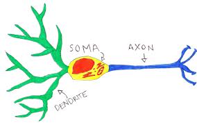
The purpose of this interactive program is engage in authentic scientific discovery related to synaptic transmission in central synapses as well as at neuromuscular junctions. |
||
|
Too often life science is distilled into disparate facts addressing major biological concepts, but lacking purpose for learning and applying knowledge to real world contexts. The driving principle of this project is to to foster authentic scientific investigation in biological sciences within the field of neurobiology. |
||
|
|
||
|
Resources: Papers and web links FREE
NEUROPHYSIOLOGY TEXT BOOK ON LINE (use for a review of basic concepts) Free background information on neuron morphology, conducting electrical, ionic bases of resting & action potential, and Synapse structure Participation in live experiment in some schools might be difficult but BACKYARD BRAINS makes equipment for students to be able to use and record neurophysiological data for introducing concepts. Participation in conducting analysis of synaptic responses will be the main focus in this course. |
||
|
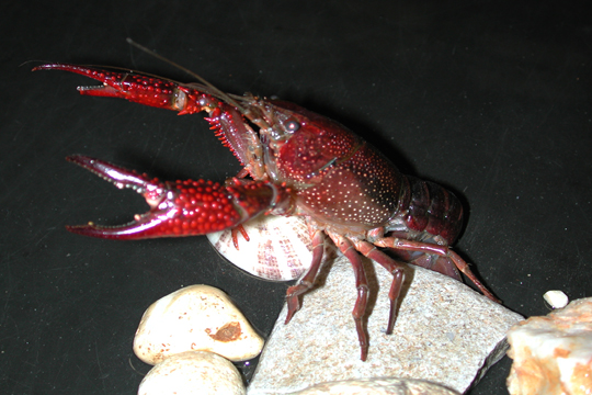
The learning objective of this laboratory is to be able observe and measure quantal synaptic vesicular release of neurotransmitter at the crayfish and Drosophila neuromuscular junctions (NMJ) as well as analyze electrical activity within the CNS of these animals. Students will participate in authentic scientific data gathering and help to improve the methods for this science project. The primary data is obtained in one lab and then posted on line for participants to download. The synaptic responses are experimentally recorded when a motor nerve is electrically stimulating for inducing evoked responses. Spontaneous vesicular events are also recorded in the absence of stimulation or between evoked stimulations. The participants estimate means quantal content with three different approaches direct counts, amplitude measurements, and charge (area) measurements. Other forms in analysis of activity from CNS circuits will also be tackled.
Data files posted: {1 mM Ca2+ in the bath} File 1: 20, 40 and 60 Hz with 30 sec wait between each trail . Each condition repeated 10 times with 10 stimuli each.. 01-17-2017 prep 1 -30 sec delays .............. DATA SHEET Excel file for 1st, 5th and 10th trials A file not for analysis but to see the evoked responses EPSPs with only 10 sec between trails EPSP views prep 2- 10 sec delays File 2: This is now of the same type of experiments but instead of 1 mM Ca2+ in the bath we only have 0.1 mM Ca2+. The 1st part of the file shows the change from 1 mM to 0.1 mM so you can see the effect on the evoked release. Then we go to high gain to examine minis at 20 Hz, 40 Hz and 60 Hz stimulations of 10 pulses with 30 sec intervals for the 10 trials for each condition. The file 0'1mmCa 30 second interval 1-23-17.adicht is name as such since saving the file as 0.1 some times causes windows machines to go crazy with the " . " File 3: This file is with 20 stimulations within a train. The frequencies are the same as for the 10 stimultions within a train (20 Hz, 40 Hz and 60 Hz) with 30 seconds between trains. Again lets only analyize the frequncy of minis after the 1st, 5th and 10th stimulation trains for each of the frequencies. The file is here And this is with 1.0 mM Ca2+ in the bath. File 4: D42-XXL dark adapted. CNS cut out. 10 uM ATR 24 hrs.... 4 trials with 1 sec blue light FILE here File 5: TBA >>>>>>>>>>>>>>>>>>>>>>>> PPTs: Intro on synaptic transmission;.... Intro on Channel Rhodopsins ..... quantal variation ppt Movies: Zana, Tori intro, Tori measures Research articles: Why use flies ? (PDF 1, 2, 3, 4 flies and human disease ) Minis different than evoked...... Spontaneous and Evoked Release Are Independently Abdrakhmanov, M.M., Petrov, A.M., Grigoryev, P.N., Zefirov, A.L.(2013) Depolarization-induced calcium-independent synaptic vesicle exo- and endocytosis at frog motor nerve terminals. Acta Naturae 5(4):77-82. Abstract http://www.ncbi.nlm.nih.gov/pubmed/24455186 Bradacs, H., Cooper, R.L., Msghina, M., and Atwood, H.L. (1997) Differential physiology and morphology of phasic and tonic motor axons in a crayfish limb extensor muscle. Journal of Experimental Biology 200:677-691. [FullText.pdf] Caldwell, L., Harries, P., Sydlik, S. and Schwiening, C.J. (2013) Presynaptic pH and Vesicle Fusion in Drosophila Larvae Neurones.SYNAPSE 67:729–740. [FullText.pdf] Cooper, A.S., and Cooper, R.L. (2009) Historical view and demonstration of physiology at the NMJ at the crayfish opener muscle. Journal of Visualized Experiments (JoVE). JoVE. 33. http://www.jove.com/index/details.stp?id=1595; doi: 10.3791/1595. Text part of video article [PDF]. Cooper, R.L., Harrington, C. Marin, L., and Atwood, H.L. (1996) Quantal release at visualized terminals of crayfish motor axon: Intraterminal and regional differences. Journal of Comparative Neurology 375:583-600 [Abstract] [Full text PDF] Cooper, R.L., Marin, L., and Atwood, H.L. (1995) Synaptic differentiation of a single motor neuron: conjoint definition of transmitter release, presynaptic calcium signals, and ultrastructure. Journal of Neuroscience 15:4209-4222 [Abstract] [pdf] Cooper, R.L., Stewart, B.A., Wojtowicz, J.M., Wang, S., and Atwood, H.L. (1995) Quantal measurement and analysis methods compared for crayfish and Drosophila neuromuscular junctions and rat hippocampus. Journal of Neuroscience Methods 61:67-78 [Abstract] [PDF] Cooper, R.L. and Ruffner, M.E. (1998) Depression of synaptic efficacy at intermolt in crayfish neuromuscular junctions by 20-Hydroxyecdysone, a molting hormone. Journal of Neurophysiology 79:1931-1941 [Abstract] [FullText.pdf] Lancaster, M., Viele, K., Johnstone, A.F.M., and Cooper, R.L. (2007) Automated classification of evoked quantal events. Journal of Neuroscience Methods 159: 325-336. [PDF] Lee, J.-Y., Bhatt, D., Bhatt, D., Chung, W.-Y., and Cooper, R.L. (2009) Furthering pharmacological and physiological assessment of the glutamatergic receptors at the Drosophila neuromuscular junction. Comparative Biochemistry and Physiology, Part C 150: 546–557[PDF]. Li, H. , Peng, X., and Cooper, R.L. (2002) Development of Drosophila larval neuromuscular junctions: Maintaining synaptic strength. Neuroscience 115:505-513 [PDF] Thieffry M. (1984) The effect of calcium ions on the glutamate response and its desensitization in crayfish muscle fibres. J. Physiol. 355:119-135. [PDF] Viele, K., Stromberg, A., and Cooper, R.L. (2003) Determining the number of release sites within the nerve terminal by statistical analysis of synaptic current characteristics. Synapse 47:15-25 [PDF] Wu, W.H. and Cooper, R.L. (2010) Physiological recordings of high and low output NMJs on the Crayfish leg extensor muscle. Journal of Visualized Experiments (JoVE). Jove 45: http://www.jove.com/index/details.stp?id=2319 , doi:10.3791/2319 [PDF of paper] Wu, W.-H. and Cooper, R.L. (2012) The regulation and packaging of synaptic vesicles as related to recruitment within glutamatergic synapses. Neuroscience 225:185-198. [PDF] Wu, W.-H. and Cooper, R.L. (2013) Physiological separation of vesicle pools in low- and high-output nerve terminals. Neuroscience Research 75: 275–282. [PDF] |
||
|
|
||
|
website
maintained by Robin L. Cooper. Contact: RLCOOP1 at UKY.EDU |